History of Spatial Transcriptomics: Spatial Transcriptomics of Tissue Regions

To overcome the limitation of only being able to study gene expression in areas of a tissue that were isolated by LCM, a group at The Spatial Research Lab at the Science for Life Laboratory developed a novel slide-based transcriptomics approach. This group was led by Drs. Joakim Lundeberg, Stefania Giacomello, and Patrik Ståhl (14). The spatial transcriptomics (ST) technology they developed utilized single-stranded DNA probes, spotted on a slide in a manner similar to microarrays. These slide-bound DNA oligo probes contain a spatial barcode to map back to its position on the slide for spatial expression analysis. The oligo also contains a Unique Molecular Identifier (UMI) to differentiate the probes on the same spot. At the 3’ end of the oligo probe, it contains a poly-T tail to capture polyadenylated mRNA released from the permeabilized tissue. The captured mRNA is reverse transcribed resulting in a spatially barcoded cDNA that is prepared as a library for next generation sequencing (NGS) (14). The end result is whole transcriptome sequencing data that can be mapped back to its original location in the tissue. Initially, the ST microarray consisted of ~1000 spots, each with a diameter of 100um. At the end of 2019, the ST technology was acquired by 10X Genomics and commercialized under the name Visium. 10X Genomics increased the number of spots on the array to ~5000 and reduced the diameter of each spot to 55um. At this resolution, each spot covers 1-10 cells. Although this greatly improved the resolution for ST, it is still not able reach the level of single cell or the ultimate goal of subcellular resolution.

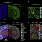
In 2019, NanoString Technologies also launched its first spatial transcriptomics platform, GeoMx DSP (Digital Spatial Profiler). This technology enabled multiplexed spatial profiling of not only RNA but also proteins. Unlike Visium’s method of capturing and sequencing RNA via an oligo dT probe, the GeoMx DSP probe uses a unique complementary sequence to directly hybridize to a transcript of interest. This target-specific capture probe is attached to a DSP barcode via a photocleavable linker. The DSP barcode is used to backtrack the location of the transcript in the tissue. These barcodes can be analyzed downstream in the workflow using NanoString’s “n-counter” system or by NGS, depending on the number of genes desired for analysis (~1000 or ~18,000, respectively). Two of the most significant limitations of the GeoMx DSP system are its resolution and single molecule detection approach. Like Visium, GeoMx cannot detect genes expressed in individual cells (resolution can only go down to 100µm).

Top questions about spatial transcriptomics

What is a Unique Molecular Identifier (UMI) for Stereo-seq?

Unique Molecular Identifiers or UMI are short sequences or molecular “tags” contained in each oligo on a Stereo-seq chip. Each oligo within a spot on the Stereo-seq chip has its own UMI to identify that individual oligo.

What are limitations of the Visium and GeoMx Digital Spatial Profiler technologies?

Two of the most significant limitations of the GeoMx DSP system are its resolution and high cost. Both 10x Genomic’s Visium and NanoString’s GeoMx DSP cannot detect gene expression at the single cell level as each spot typically overlaps 5 to 10 cells. The costs to run each of these platforms, including sequencing, is often over $10,000 for each sample.


History of Spatial Transcriptomics: Single Gene Spatial Transcriptomics

The function of complex multicellular organisms depends on the spatial organization of their cells. It is accepted that all cells in an organism (except for germ cells and lymphocytes ) have the same genetic make-up. Then, what contributes to diversity of cells and their function? Mostly, differential gene expression. Thus, the study of the spatial patterns of gene expression in complex tissues has been used to answer fundamental questions in several scientific disciplines such as developmental biology, neuroscience, cancer biology, and pathology (1–4).

Initially, these spatial expression studies looked at single genes. One of the earliest methods of detecting spatial gene expression was in situ hybridization (ISH), used by Walter Gehring’s group in the fly (Drosophila) embryo in the 1980’s. Their goal was to characterize key genes and their spatial expression during the Drosophila embryo segmentation process (5). Gehring’s group focused on the fushi tarazu (ftz) gene. By using ISH, they found that the ftz transcript is expressed in a seven-striped pattern. When ftz is mutated, this results in missing alternating segments. In subsequent studies, by independent labs, multiple genes were shown to be involved in Drosophila’s embryo segmentation (6). Even though these studies were pioneering at the time, the technology available limited the study of expression patterns to one gene at a time.

A significant advancement in spatial transcriptomics was made with the development of laser capture microdissection (LCM) (7). Using LCM, one can dissect a small region of a complex, heterogeneous tissue for deeper study. In the early days of LCM, gene expression analysis was typically done using northern blotting or RT-PCR (8,9). However, these techniques still limited the number of genes studied. This limitation was overcome in 1997 with the arrival of high-density DNA microarrays, which allowed for the first time to analyze over 2000 genes in yeast (10). In the early 2000s, these microarrays were used to analyze LCM samples, greatly expanding the number of genes to study. Early experiments utilized LCM tissues from brain, breast, and prostate tumor tissues for gene expression profiling using microarrays By around 2010, microarrays were slowly being replaced by RNA-sequencing technologies. With this technique, we can capture expression of most existing transcripts. However, even with the advent of RNAseq, LCM still has its limitations in how small of a region can be example. Anything smaller than 30 µm in diameter risks collateral cellular damage resulting from cutting techniques (laser vs UV). Another disadvantage of LCM based methods of studying gene expression is that it is limited to characterizing the area isolated, leaving out surrounding areas that are needed to understand spatial biology.


Top questions about spatial transcriptomics

Who was an early pioneer of spatial transcriptomics?

Walter Gehring was one of the fathers of modern developmental biology. In the 1980’s, he became one of the first to isolate, identify, and characterize developmental control genes 1980’s. To accomplish this feat, Gehring and his collaborators pioneered a technique called in situ hybridization (ISH). With this technique, a biological sample is affixed to a glass slide and then exposed to a probe consisting of a small piece of single-stranded nucleotide sequence that is complementary to a target of interest. This probe is tagged with a chemical or fluorescent dye. ISH was one of the earliest methods of detecting spatial gene expression. It was used widely across most model organisms for biology research and is still being used today.

What is laser capture microdissection (LCM)?

Laser-capture microdissection (LCM), also known as Laser Microdissection (LMD) is a method for isolating cellular regions of tissue under direct microscopic visualization. LCM technology can be used to isolate specific groups of cells from a mixed population of cells. By characterizing these subpopulations of cells, one can examine the cellular and molecular interactions that drive disease within the tissue microenvironment. Understanding these cellular and molecular interactions can be used for identify drug targets for treatment.